Imagine this: you’ve just completed your multiplex immunofluorescence experiment, and when...
Introducing CellScape™ Whole-Slide Imaging Chamber
Now you can convert any standard microscope slide into a microfluidic chamber for automated multiplex staining, high-resolution imaging, and safe sample archiving. Read more about the expansion of our Precise Spatial Multiplexing capabilities.
Whole-slide spatial biology
Spatial biology is a rapidly growing field that enables exploration and visualization of tissues for a wide variety of applications, including basic research, drug discovery, diagnostic development, and treatment monitoring. Imaging large tissue sections or several samples on the same slide, such as tissue microarrays (TMAs), with whole-slide spatial techniques offers numerous benefits:
- More comprehensive analyses- imaging large areas increases the likelihood of capturing key biological processes.
- Expanded throughput- imaging multiple samples on the same slide enables broader data acquisition at reduced reagent costs.
- Improved statistical power- placing multiple samples on a single slide increases sample numbers, supporting more statistically significant conclusions.
- Enhanced value for clinical applications- imaging larger samples increases the chance of detecting clinically-relevant anomalies in tissues.
- Tissue architectural analysis- whole-slide imaging enables more complex analyses of biological processes in relation to larger tissue structures.
CellScape™ Precise Spatial Multiplexing for highly multiplexed biomarker detection
The CellScape platform enables automated cyclic multiplex immunofluorescence imaging with unprecedented optical resolution needed for quantitative spatial phenotyping. The most versatile instrument for spatial biology, the CellScape utilizes standard fluorescent antibodies for biomarker detection and is compatible with any species or sample type. Leveraging both high-resolution optics and high dynamic range (HDR) imaging, CellScape can detect biomarkers across a wide range of expression levels.
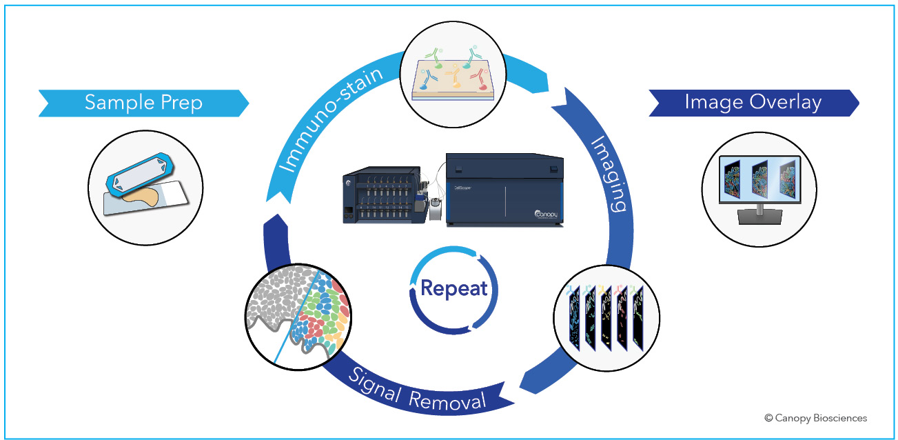
The CellScape platform employs iterative cycles of staining, imaging, and signal removal via gentle photo-inactivation to detect biomarkers with spatial context and single-cell and subcellular resolution. View the CellScape brochure to learn more about the benefits of the instrument.
CellScape Whole-Slide Imaging Chamber enables compatibility of CellScape with slides
The CellScape Whole-Slide Imaging Chamber is compatible with any standard histology microscope slide (25 mm by 75 mm). The chamber is composed of a glass coverslip with a removable adhesive on one side. Features of an assembled Slide with Imaging Chamber include:
- 710 mm2 available imaging area
- 50 µL imaging chamber volume
- Automated reagent delivery through imaging chamber inlet and outlet
- Uniform reagent delivery via integrated flow diverters
- Long-term biobanking capability
- Unique sample identification via QR and human readable codes
- Archived FFPE sample compatibility across any species
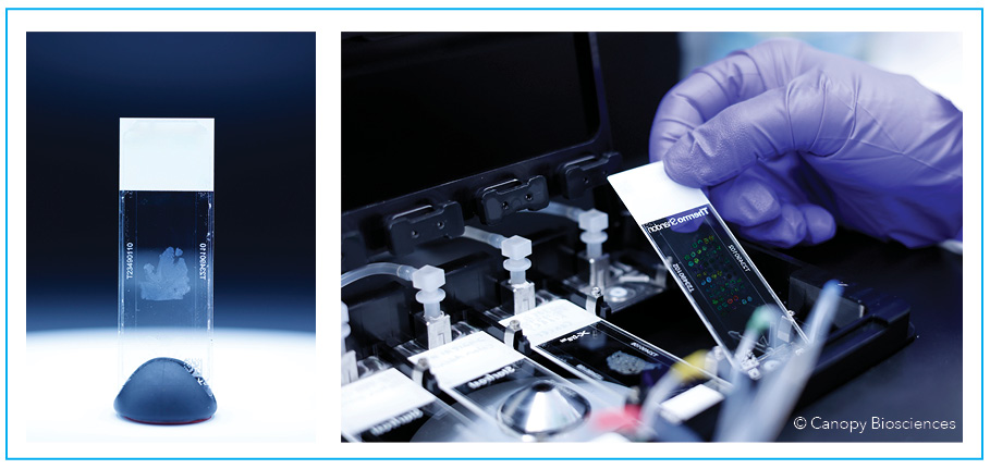
Assembled CellScape Whole-Slide Imaging Chamber (left) and assembled slides being loaded into CellScape instrument (right).
Preparation of a Whole-Slide Imaging Chamber
Assembly of the chamber with a slide is simple with the CellScape Whole-Slide Assembly Tool. The tool enables precise alignment, optimizes imaging window placement, and standardizes sample prep for reproducible and reliable results.
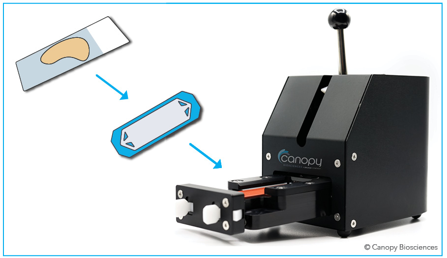
CellScape Whole-Slide Assembly Tool aligns a slide with the CellScape Whole-Slide Imaging Chamber and creates a seal with the necessary amount of uniform pressure, avoiding misalignment, incomplete sealing, and cracking of glass.
Cost reduction using CellScape Whole-Slide Imaging Chamber
In addition to providing the maximal viewing area, the CellScape Whole-Slide Imaging Chamber saves costs on reagents compared to other technologies. The platform is compatible with standard, off-the-shelf fluorescent antibodies that are utilized at dilute concentrations, so there is no requirement for custom antibody conjugations or reagent modifications. Additionally, the Whole-Slide Imaging Chamber uses small volumes of reagents, providing an advancement to the original ChipCytometry™ technology utilized by the CellScape platform. The opportunity to combine multiple samples on a single slide further decreases the cost per sample while increasing throughput.
Biobanking and data-driven assay expansion using CellScape Whole-Slide Imaging Chambers
A unique feature enabled by CellScape is that samples prepared in Whole-Slide Imaging Chambers are capable of storage and reexamination—follow-up staining and imaging of an already-analyzed sample. This data-driven assay expansion provides a flexible and modular approach to high-plex spatial biology. Benefits of assay expansion include:
- On-the-go assay development
- Online quality control of tissue specimens and evaluation of samples for assay suitability
- Ad-hoc assay expansion
- Progressive troubleshooting
- Conservation of precious or rare samples
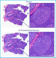
Below, data-driven assay expansion with a CellScape Whole-Slide Imaging Chamber was demonstrated using two VistaPlex™ Assay Kits, modular and efficient panels for detecting common biomarkers using a CellScape instrument.
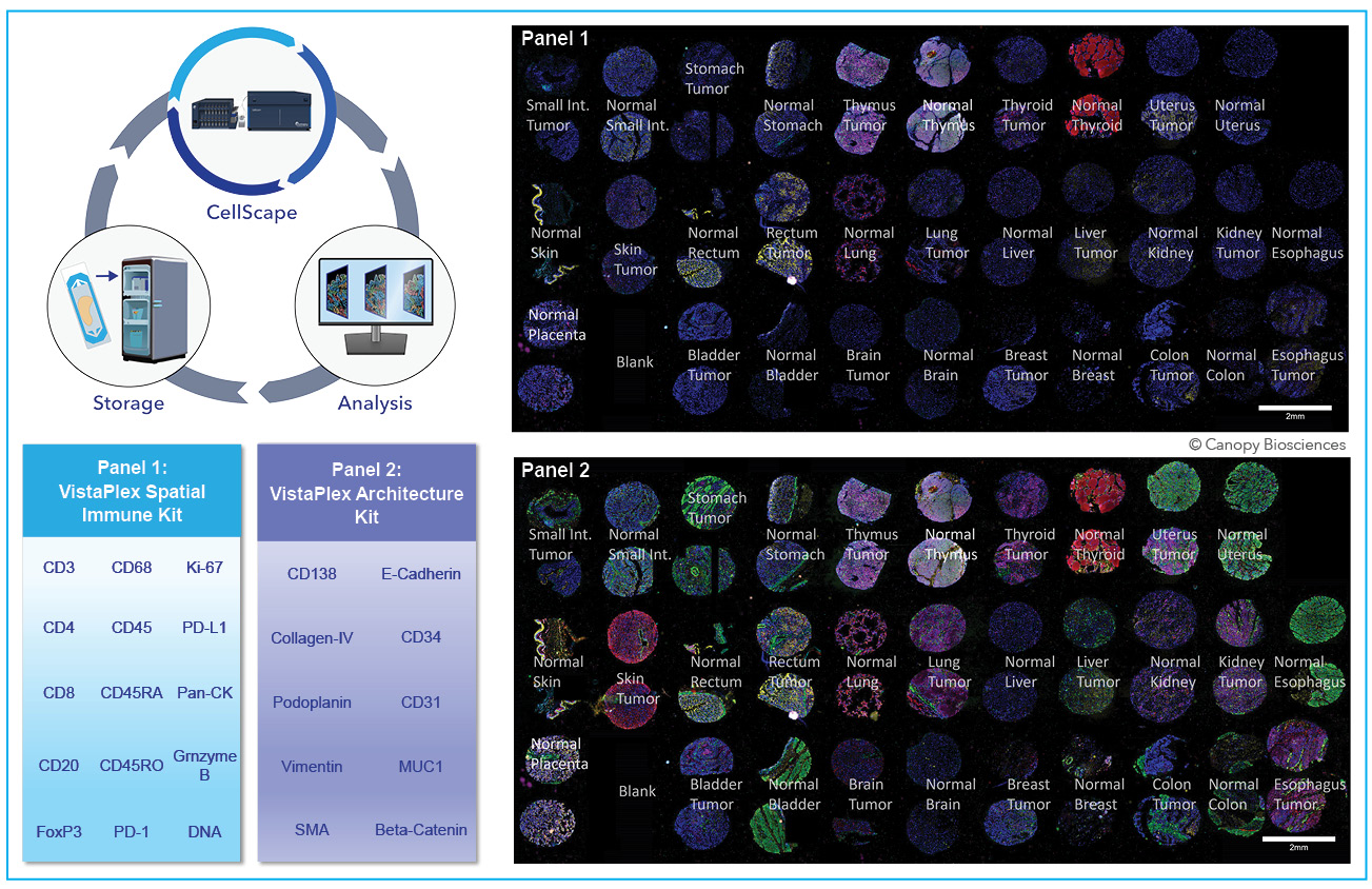
Data-driven assay expansion workflow using CellScape includes iterative automated staining and imaging followed by data analysis and sample storage. The nature of the CellScape workflow enables the user to pause between rounds of staining/imaging and determine how (or even whether) to continue to the experiment.
To learn more about data-driven assay expansion using CellScape, read our Application Note.
Access to Whole-Slide Imaging for existing CellScape users
Current CellScape users will have the opportunity to upgrade their instrument to be compatible with the new Whole-Slide Imaging Chamber. The field upgrade is completed by replacing the stage insert. For more information or to schedule an upgrade now, please fill out this form.
Achieve best-in-class spatial biology data with Cellscape Whole-Slide Imaging
The CellScape platform enables high-plex spatial omics with quantitative readouts and flexible reagent choices. Now, the improved sample preparation workflow expands the platform’s capabilities even further by supporting spatial biology without limits, unlocking whole-slide exploration and powerful sample data-driven assay expansion.
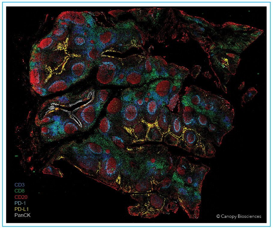
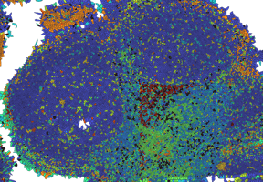
High resolution image of FFPE human tonsil stained with 14-plex VistaPlex Spatial Immune Profiling Assay Kit, imaged with CellScape Whole-Slide Imaging Chamber. Select biomarkers are displayed as indicated in the legend. The increased scanning area available with Whole-Slide Imaging enables comprehensive analysis of large tissue samples.
Ready to get started? Contact us today for more information or a quote for your new CellScape.