Spatial Biology: What it is, why it matters, how to access it.
Why You Need to Increase the Throughput of Your Single-Cell Imaging Experiments
Is your world filled with terms like spatial biology, single-cell analysis, and multiplexed imaging? With a variety of single-cell imaging techniques on the market, it can be time consuming to thoroughly investigate which instrument or CRO partner is right for your research. Canopy Biosciences breaks down several factors to consider when deciding which instrument is right for your high-throughput single-cell imaging experiments.
What is high-throughput single-cell imaging?
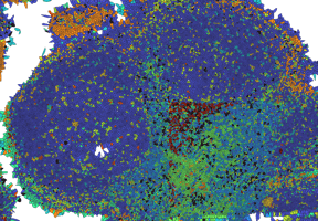
High-throughput single-cell imaging involves using automated microscopes to acquire images of individual cells at superior resolution within a short period of time. This technique has recently exploded as researchers recognize its utility to generate a significant amount of data and uncover new biological insights at unprecedented detail. High-throughput single-cell imaging is particularly useful because it allows researchers to study heterogeneity and cell diversity within a single biological specimen. This feature is particularly important in tissues, where a single specimen can contain a wide variety of cell types organized in epithelial, immune, and stromal compartments within the tissue. High-throughput single-cell imaging is useful to study cell-cell interactions, tissue microenvironment, and immune response to therapeutics across a range of applications in oncology, immuno-oncology, and neuroscience.
Why is high-throughput single-cell imaging important?
High-throughput single-cell imaging has enabled researchers to collect more data in a shorter amount of time, without compromising image quality. Researchers can now collect more information than ever before on thousands of single cells within a single biological specimen, including cell phenotype, cell state, and spatial position. The key feature of high-throughput single-cell imaging is that it allows researchers to deeply phenotype cells in situ to maintain important information about the spatial relationship between cells. With a variety of single-cell imaging techniques on the market, there are many factors to consider when deciding which instrument is right for your research. Specifically, instruments that maximize throughput without compromising the resolution necessary to achieve single-cell detail or plex necessary to deeply profile specimens. Instruments designed with throughput in mind and CRO partners with quick turn-around-times, will lead the way in this regard.
What factors influence the throughput of single-cell imaging?
There are many factors that influence the throughput of a single-cell imaging experiment. It is important to consider factors related to both instrument and experiment design:
- The resolution of images: Higher resolution images take longer to collect and therefore decrease throughput.
- Total scanning area: A larger scanning area decreases throughput by increasing the total imaging time.
- The number of targets: Higher plex assays decrease throughput by increasing the total imaging time.
- The number of channels: More imaging channels decreases throughput by increasing the total imaging time.
- The size of the field-of-view (FOV): A larger FOV increases throughput by allowing more cells to be imaged at once.
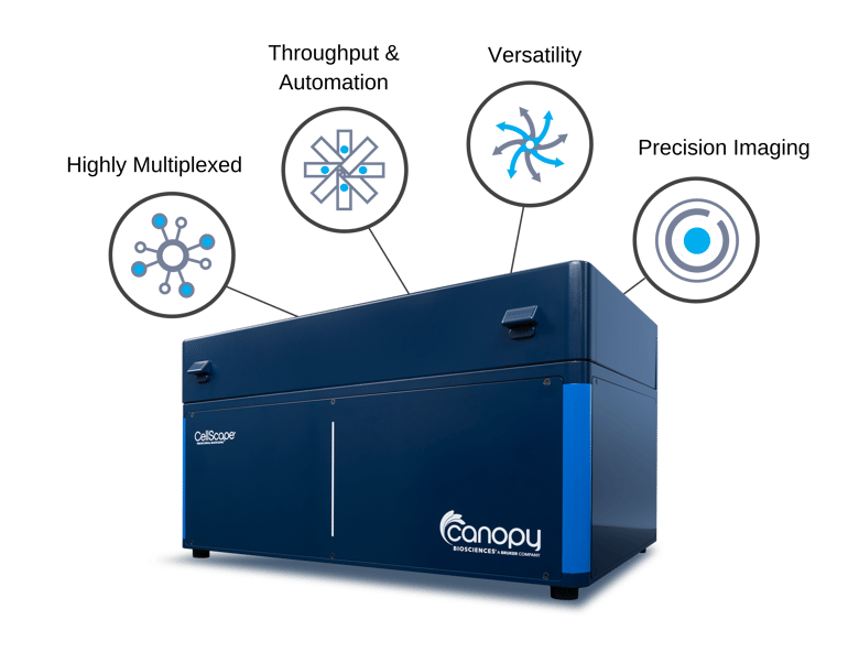
Although maximizing throughput may seem like a function of simply optimizing each of these features independently, the reality is far more complex. Researchers want it all: resolution and plex without compromising on throughput. The CellScape™ instrument from Canopy Biosciences, which comes in an optional FalconFAST mode, was designed with this in mind and combines several unique features to maximize throughput without compromising resolution or plex.
How does CellScape compare with other instruments?
CellScape combines several unique features including (i) 5-color imaging and (ii) large field-of-views (FOVs) for higher throughput experiments. The 5-color imaging refers to the number of fluorescence channels in the CellScape instrument. In fluorescence microscopy, spectral overlap from fluorophores makes it challenging to isolate signal from specific targets [1]. Therefore, the number of channels in any fluorescence microscope is limited to around 5-6 to be able to spectrally resolve fluorophores. More channels are better, as long as fluorescent signals can be isolated. Especially for instruments with cyclic workflows, having more imaging channels is advantageous to reduce the total number of cycles, each of which involve a 10–60-minute antibody staining and incubation step. Another key feature of CellScape is large field-of-views (FOVs) which allow for many more cells to be imaged at a time. FOV size is also largely determined by instrument design, specifically the optical components and camera. FOV size is often an overlooked feature, which should be given more attention because it significant affects experiment throughput. These features, when combined, are unique to the CellScape instrument and support higher throughput single-cell imaging experiments.
What is the throughput of CellScape?
With CellScape, it is possible to image 4-8 tissue sections in 24 hours using a standard 20-plex assay, with ~10 mm2 scanning area. The best way to increase throughput with CellScape is with FalconFAST mode, which reduces total imaging time for studies that require ultrahigh-throughput imaging. With FalconFAST mode, it is possible to image 8 samples in 24 hours using a standard 20-plex ChipCytometry™ assay. As with any single-cell imaging experiment, the actual throughput depends on experiment design.
How does experiment design affect throughput?
Throughput is also affected by scanning area, number of markers, and panel design. In some experiments, it is necessary to image the entire tissue section to capture cell heterogeneity across the whole slide. As in any single-cell imaging experiment, this will increase image acquisition time and decrease throughput. Similarly, some studies require ultrahigh-plex assays where the number of makers in an antibody panel exceeds 40-50 markers. An increase in the number of targets will also increase image acquisition time and decrease throughput. However, throughput can be improved with smart panel design. Thoughtfully structured antibody panels fill each channel in a cycle to minimize the total number of cycles. In some cases, this may mean selecting an antibody conjugated to a different fluorophore to move it to a channel in an early cycle. The brilliance about CellScape and the ChipCytometry workflow is that it is flexible enough to allow this.
For Research Use Only. Not for use in diagnostic procedures.
References
[1] Neher, R., & Neher, E. (2004). Optimizing imaging parameters for the separation of multiple labels in a fluorescence image. Journal of Microscopy, 213(1), 46–62. https://doi.org/10.1111/j.1365-2818.2004.01262.x